Introduction
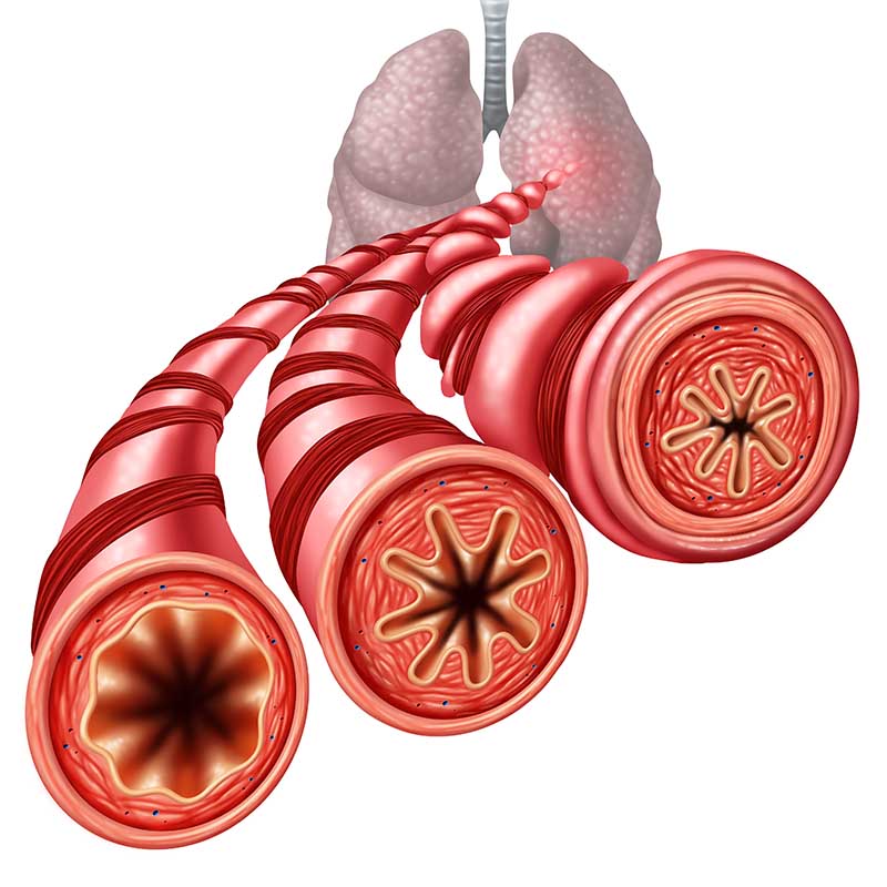
Acute flank pain secondary to obstructive ureteric stone is often associated with visits to the emergency department (ED). Noncontrast computed tomography (NCCT) is the first line treatment but associated with higher cost and exposure to ionizing radiations. Ultrasound (US) is excellent for detection of hydronephrosis but has a poor sensitivity for stone detection. Radiography of kidney-ureter-bladder has better sensitivity for stone detection but poor sensitivity for hydronephrosis detection. A combination of both these modalities might improve the sensitivity and specificity for the diagnosis of obstructive ureteric stone and hydronephrosis. However, there is paucity of studies which have evaluated the utility of combining both these modalities in the same patients.
Aim
The study evaluated the diagnostic accuracy of combined US and radiography for the diagnosis of obstructive ureteric stone in adult patients.
Method
Study Design
- Retrospective study
Treatment Strategy
- A total of 164 adult patients presenting with acute flank pain to the ED who received both US and radiography at the ED were enrolled
- Study period- August 1, 2014, to July 31, 2015
- The decision for NCCT was based on US and radiographic findings and a clinical probability stratification called STONE score
- The patients were stratified based on the STONE score into three groups of low, intermediate, and high probability of ureteric stone.
- The patient was managed according to STONE score and initial ultrasound/radiographic findings
- A total of 90 patients (45 men, mean age 45.6 years, range 16–90 years) presenting with acute flank pain underwent NCCT and constituted final cohort
- The clinical reports, pathological, radiological and laboratory tests, electronic medical records were analyzed
- The computed tomography was used as diagnostic reference
Endpoints
- Diagnostic performance of US, radiography and combined US and radiography
- Sensitivity
- Specificity
- Positive predictive value (PPV)
- Negative predictive value (NPV)
- Accuracy
Results
- The STONE scores were low, intermediate, and high in 12, 51, and 27 patients, respectively
- Obstructive ureteric stone was diagnosed by NCCT in 54%
- Stone size was less than 5 mm in size in 83.7%
- The diagnostic performance of the modalities was compared using NCCT as the reference standard as can be seen in table 1.
| Modality | Diagnosis | Sensitivity (%) | Specificity(%) | PPV(%) | NPV (%) | Diagnostic odds ratio | Accuracy (%) |
| US | Hydronephrosis | 73.5 | 92.7 | 92.3 | 74.5 | 35.1 | 82 |
| US | Hydroneprosis and Ureteric stone | 14.3 | 100 | 100 | 49.4 | NA | 53 |
| Radiography | Ureteric stone | 34.7 | 100 | 100 | 56.2 | NA | 64 |
| Combined US with radiography | Hydronephrosis and ureteric stone | 87.8 | 92.7 | 93.5 | 86.4 | 90.8 | 90 |
Conclusion
- Combined ultrasound (US) with radiography offers promising result for the diagnosis of obstructive ureteric stone in patients presenting with acute flank pain to the emergency department
- The combined modality approach might reduce the need for computed tomography (CT) scan, thereby reducing the radiation exposure to the patient.
J Med Ultrasound. 2020 Apr-Jun; 28(2): 86–91.