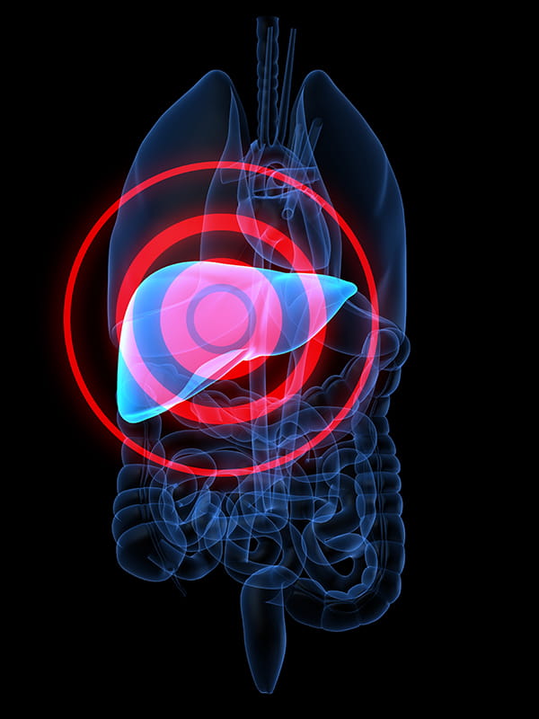
DDW 2024: Evaluation of Liver Disease in the Community


The session discussed making accurate diagnosis and assessing of fibrosis stage, ideally in non-invasively in the community setting. Once an accurate diagnosis is made and a fibrosis stage is known the therapy can be directed to prevent progression of disease, prevent cirrhosis and its complications.
The presenter discussed the case of a 58 year old female referred to the gastroenterology center from her primary care provider for chronically elevated liver enzymes. Her recent lab reports showed a normal blood count and chemistries but her alanine transaminase (ALT) was mildly elevated (85) and aspartate aminotransferase (AST) were 55. Her alkaline phosphatase, bilirubin, albumin, INR and AFP were normal. She had diabetes, hyperlipidemia, obesity and irritable bowel syndrome. She was prescribed metformin, tylenol and dicyclomine; she did not consume alcohol, smoke or take any illicit drugs now or in the past. Her father passed away from cirrhosis secondary to alcohol associated liver disease. In this case, the first step was to make an accurate diagnosis of seeing a patient with elevated liver enzymes. Detailed diagnosis revealed that she had paracellular type injury. She had negative hepatitis B surface antigen and hepatitis C antibody. Her ceruloplasmin was normal (24), ferritin was slightly elevated at 388 with a normal iron saturation, alpha 1 antitrypsin level was normal and her phenotype was normal as well. She had a mildly positive ANA at 1:40 but a negative anti-smooth muscle antibody and a normal IgG level. Imaging studies revealed hepatomegaly increased echogenicity consistent with hepatic steatosis. There were no masses, and her spleen was normal in size. From this information many causes of chronic liver disease can be ruled out and she does not have alcohol associated liver disease.
She did not have drug induced liver injury, viral hepatitis, Wilson's disease or alpha 1 antitrypsin deficiency. However, there is a suspicion of metabolic dysfunction associated steatotic liver disease. Since her ferritin levels were, the diagnostic challenge was that is this hemochromatosis? The AASLD guidelines recommend conducting an HFE genotyping anyone with an iron saturation higher than 45% and/or an elevated ferritin. It is important to keep in mind that having a chronic liver disease, especially non-alcoholic fatty liver disease (NAFLD), is associated with iron overload and with elevated ferritin levels additionally related to inflammation. The second diagnostic challenge was autoimmune hepatitis. At the tertiary level a number of people have been mislabeled as fatty liver when autoimmune hepatitis was the underlying diagnosis. Autoimmune hepatitis requires a serologic positive autoantibodies and confirmatory histopathology. Additionally, multiple other chronic liver diseases are associated with positive autoantibodies. In 34% of patients with NAFLD, the ANA will be positive. In the case described above, she had positive ANA, negative anti smooth muscle, a normal immunoglobulin level and conventional risk factors for NAFLD. ruling out autoimmune hepatitis. The ASLD website has a decision tree to help one in on the diagnosis of MASLD. The decision tree involves yes or no questions to know whether the patient has elevated liver enzymes or whether they have steatosis on imaging. With this decision, the case under discussion suffers from MASLD.
The second step in her assessment that helps us manage and therefore prevent problems is staging her liver disease. Patients with MASLD who have higher than F2 fibrosis are the patients that are likely to progress and have liver related events. Patients that have earlier stages of MASLD have higher mortality from cardiovascular causes as well as non-hepatic malignancies. There are two scoring systems that are most commonly used which require basic blood work in a patient, age and weight. The FIB 4 score uses age, AST, platelets and ALT to give a score. If the score is <1.3, then you exclude significant fibrosis. The NAFLD fibrosis score includes age, BMI, the presence of insulin resistance for diabetes, liver enzymes, platelets and albumin. A score of <-1.455 excludes fibrosis with good test characteristics. The Enhanced Liver Fibrosis (ELF) test uses 3 serum biomarkers and combines it into a score which is meant to assess the risk of disease progression. If the score is <9.8, then the patient is at low risk of progression. If it is >11.3, they are at a higher risk of progression and you'll see where this comes into play with an algorithm and a couple of slides. Transient elastography or Fibroscan gives a liver stiffness measurement measured in Kilopascal. A measurement <8 rules out advanced fibrosis and >12 indicates a likelihood of advanced fibrosis. Ultrasound elastography and MR elastography are some of the other imaging techniques to diagnose fibrosis. According to the ASLD guidelines, patients with high fibrosis score indicating an advanced stage are guided to the gastrointestinal/hepatology care. Patients with FIB 4 or secondary stage may benefit from a retest such as a Fibroscan. In absence of a Fibroscan, the ELF test can be used. If the tests are inconclusive, a liver biopsy may be considered. In case of an early-stage disease, the patient can be reassessed for fibrosis in 2 to 3 years. In stage 2 to 3, pharmacotherapy can be considered and reassess the patients annually.
There are 3 new practice guidelines that are available through AASLD which discuss blood based and imaging based non-invasive fibrosis and steatosis testing. They also help with the determination of whether someone has portal hypertension. In a quick summary, for a blood-based test, only hepatitis C has good data to support using before upfront. All other liver diseases do not have enough evidence to recommend routinely doing a FIB 4 or other serum-based tests. The imaging-based tests are better. The AASLD recommends performing imaging-based tests for initial fibrosis staging over blood-based tests for diseases other than MASLD. it is important to identify portal hypertension by screening for oesophageal viruses and adding carvedilol if there is any clinically significant portal hypertension to prevent decompensation events and their bleeding. It is encouraged to perform an ultrasound, an AFP every six months for hepatocellular carcinoma surveillance.
In conclusion, making an accurate diagnosis of chronic liver disease allows for appropriate management to prevent progression of disease. Non-invasive blood based and imaging-based tests can help determine a fibrosis stage that can help guide drug therapy and allow initiate appropriate cirrhosis surveillance.
Digestive Disease Week (DDW) 2024, May 18-21, 2024, Washington, D.C.